Clear vision today is one thing. Ensuring you have it tomorrow is something entirely different.
Advanced technology has changed the way we practise optometry, and we’ve invested heavily in instruments that provide us with the peace of mind that we want from an eye test.
Here is an example of one of these instruments, and why we use it.
Optical Coherence Tomography (OCT)
If you want to know if you have glaucoma or macular degeneration, we strongly advise you to be scanned with an OCT.
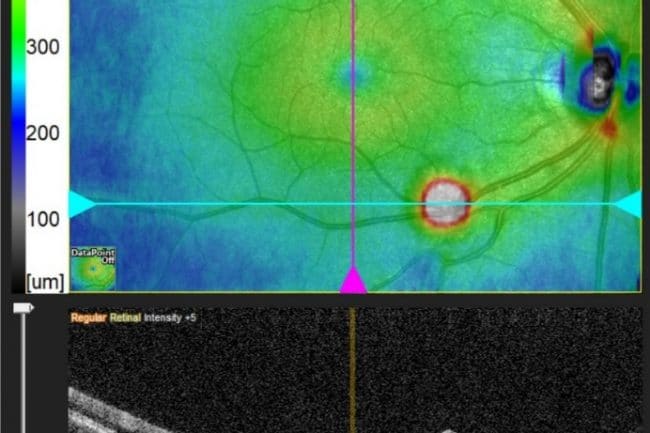
Glaucoma is the wasting away of the retina, the layer that lines the inside of the back of the eye, which leads to blindness if untreated. An OCT measures the thickness and health of the retina, detecting eye disease much earlier than conventional eye tests. And as you’re no doubt well aware, early intervention saves sight.
A case example.
There’s a lot on the net about how sophisticated these instruments are, but there’s nothing like a real-life example to explain why we recommend everyone over 40 years of age has this test every 2 years.
The case changed the way we use our OCT.
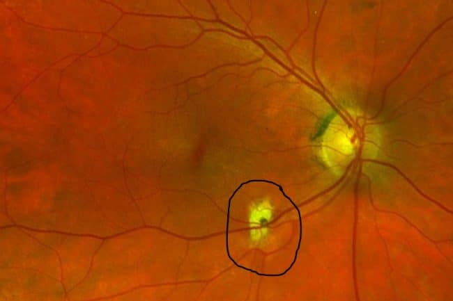
Patient X came in for a routine eye exam, with no complaints about their vision and they had no family history of glaucoma. There was no change to their glasses and the eye pressures were within the normal range. The optic nerve head looked normal, and in the past, this person would have passed as a healthy pair of eyes and left the practice after completing what’s considered a routine eye exam.
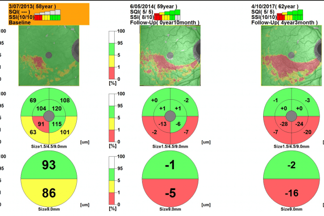
However, because patient X was over 40yrs, we scanned for glaucoma with our OCT and found the following results –
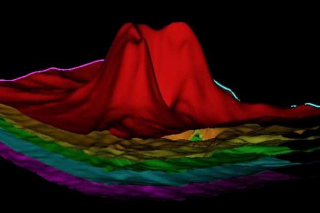
These maps show the changes in the thickness of the retina over a 4 year period: Green is good, Yellow is thinner than usual, and Red is extremely thin.
The scan above on the left raised our suspicion and a review was scheduled for 6 months. The second scan confirmed our suspicion and the person was advised to see an ophthalmologist but unfortunately didn’t listen to our advice until they noticed parts of their vision were missing. They had no pain or blurry vision to warn them, and this is exactly what glaucoma does – it silently erodes the inside of the eye, causing blindness that can’t be reversed. Yet it can be arrested if caught early.
So now you can appreciate why we no longer scan only those with a family history or suspicious looking disks. We now scan everyone over 40.
The dry stuff.
All the scans are painless and non-invasive, without the need for the instrument to come into contact with your eye, and the images are captured in just a few seconds.
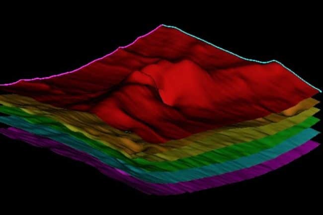
The OCT is often likened to an MRI or x-ray as it is a sophisticated scanning system that uses light waves to produce a cross-section image of the retina. And because the high resolution images can be stitched together into a 3D model, the scan is often referred to as a ‘3D Eye Scan’.
This scan allows us to see detailed images of the cornea, iris, retina and complex contact lenses that work best when they fit perfectly on the cornea. This procedure is currently the only one that shows in-depth images of the eye’s internal structures. Other procedures show only the surface of these structures.
Most people experience a change in their vision in their early 40s. Usually they notice it’s a bit more difficult to see small detail up close, like text on a phone, and this is usually caused by changes in the lens inside the eye. This is consistent with normal ageing but if glasses can’t improve the vision, it could also be a symptom of something more serious so we scan with the OCT to check your eye health. Keep in mind, up to 80% of vision impairment and blindness can be prevented by early detection.
Swollen Optic Nerve Head.
OCT is also useful in detecting disorders of the optic nerve – the bundle of retinal nerve fibres which send signals from the retina to the brain. It detects a loss of nerve fibres, a swelling of the optic nerve head or a change in the cupping inside the optic nerve head. Changes like these are caused by a range of conditions such as a brain tumour, diabetes, cardiovascular disease and glaucoma, so it’s great to have the OCT in our practice to pick them up early.
Drainage Angle Assessment.
OCT imaging of the cornea and iris assesses if there is enough room for the aqueous humour (the fluid inside the front of the eye) to efficiently drain away. Any blockages to this drainage can cause significant increase in the intraocular pressure, which might result in acute angle-closure glaucoma – a sudden, painful version of glaucoma.
Pterygium Assessment.
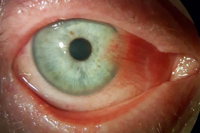
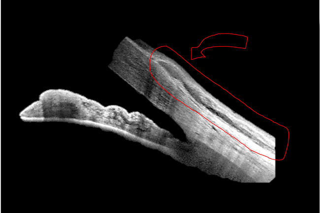
The common pterygium – the triangular skin that grows over part of the cornea near the nose like the one in the picture above – can also be assessed with the OCT. Scanning through the tip of a pterygium will determine if it penetrates into the stroma, the spongy centre of the cornea. Not all pterygia go down this far but if it does, the risk of scarring and blurred vision after surgery is much higher. So surgery in these cases should be considered earlier rather than later. This is a good time to remind anyone who spends a lot of time outdoors or works in dusty environments, that you are more likely to develop a pterygium and wearing good sunglass protection is the best way to protect your eyes.

